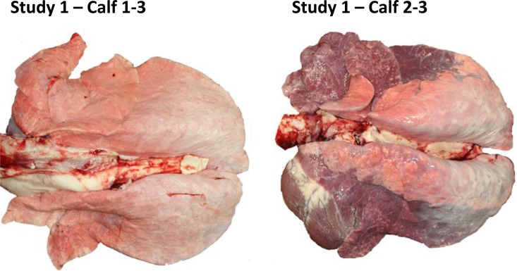
FIG 5.
Lung tissue from calves in study 1 treated with GS1 or placebo. Calf 1-3 (calf 3 from group 1) (treated with GS1) showed no gross lesions with 0% lung consolidation, with the exception of a focal fibrous adhesion on the thoracic wall. In addition, there was mild neutrophilic bronchial exudate with occasional lymphoid aggregates in bronchial and bronchiolar epithelium, and there was no active bronchopneumonia or exudation in lung. No evidence of bRSV was detected by immune staining of lung tissue. In contrast, calf 2-3 (calf 3 from group 2) (treated with placebo) showed 60% lung consolidation and widespread bronchointerstitial pneumonia with neutrophilic exudation in which bronchioles had hyperplastic epithelium with focal necrosis and surrounding lymphoplasmocytic infiltrate. Multifocal epithelial syncytia and syncytia in alveolar spaces was observed. Immune staining showed bRSV-positive signal in the lung tissue. A description of the histopathology results for individual animals is shown in Table S3 in the supplemental material.

