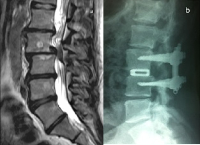
Fig. 1.
Case 15. A 41 year old man with Grade II degenerative spondylolisthesis, Level L3-L4. (a), the preoperative sagittal T2-weighted MRI illustrates the spondylolisthesis and the retropulsion of the intervertebral disc. (b), the 48-72 h postoperative sagittal radiograph shows PLIF+IPLF, with restoration of the disc space height and improvement of the spondylolisthesis.

