FIGURE 6.
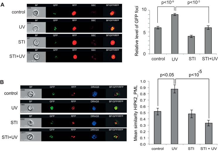
Accumulation of HIPK2 in nuclear speckles is dependent on c-Abl. A, HEK293T cells were transfected with plasmids expressing GFP-HIPK2 and RFP-histone H2B. Cells were treated with 20 μm STI571 (STI) or vehicle, as indicated, and irradiated at 50 J/m2 (UV). Cells were analyzed using the ImageStream X flow cytometer 72 h after irradiation. The left panels show bright field (BF) images, GFP-HIPK2, RFP-H2B (nuclear marker), side scatter, and merged images (BF/GFP/RFP) of representative control and UV-treated cells, with or without the c-Abl inhibitor STI571. The right panel shows quantification of GFP-HIPK2 spot count in the differently treated cell populations. Data represent S.E. from three independent experiments. The Student's t test was used to evaluate statistical significance. B, HEK293 cells were transfected with plasmids expressing GFP-HIPK2 and mRFP-PML IV. Cells were treated with 12.5 μm STI571 or vehicle, as indicated, and irradiated at 50 J/m2. Cells were analyzed as in A, but DRAQ5 staining was used to define the nucleus. The left panels show bright field (BF) images, GFP-HIPK2, RFP-PML IV, DRAQ5 (nuclear staining), and merged images (BF/GFP/RFP) of representative control and UV-treated cells, with or without STI571. Colocalization of GFP-HIPK2 and RFP-PML IV was estimated by using the Similarity Feature (Amnis) and quantified (right panel). Data represent S.E. from at least three independent experiments.

