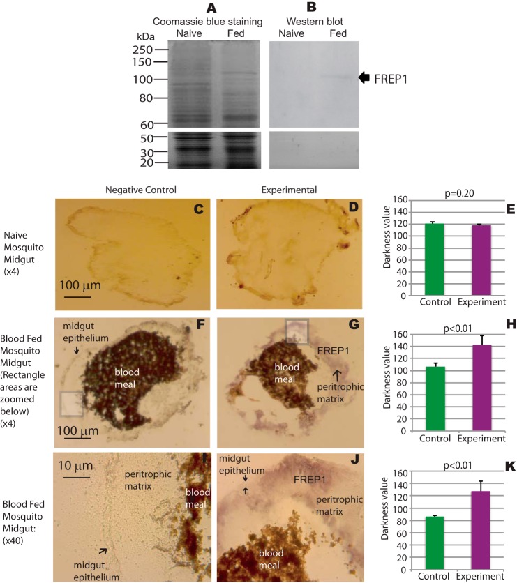
FIGURE 2.
FREP1 localizes to the mosquito midgut peritrophic matrix. A, midgut proteins from unfed (naive) and blood-fed mosquitoes were fractionated on 10% SDS-PAGE and stained with Coomassie Brilliant Blue. Approximately 10 μg of total protein, extracted from 2 to 3 midguts, was loaded per lane. B, gels were loaded as in A, and proteins were transferred to membranes and probed with 0.2 μg/ml of anti-FREP1 antibody. Western blotting data show that anti-FREP1 antibody specifically recognizes FREP1 in blood-fed mosquito midguts. C, F, and I, negative control staining of naive and blood-fed An. gambiae midgut sections using purified preimmune rabbit antibody. D, G, and J, experimental staining of naive and blood-fed An. gambiae midgut sections using anti-FREP1 rabbit antibody. FREP1 (purple signal, panels G and J) is only detected in blood-fed sections probed with anti-FREP1 antibody; I and J are ×40 magnifications of the boxed areas shown in panels F and G, respectively. Locations of the midgut epithelium, the peritrophic matrix, the FREP1, and the blood bolus are annotated on the images. Summary data (mean ± S.D.) in E, H, and K show the statistical difference of pixel intensity values between negative controls and experimental groups from three independent experiments.

