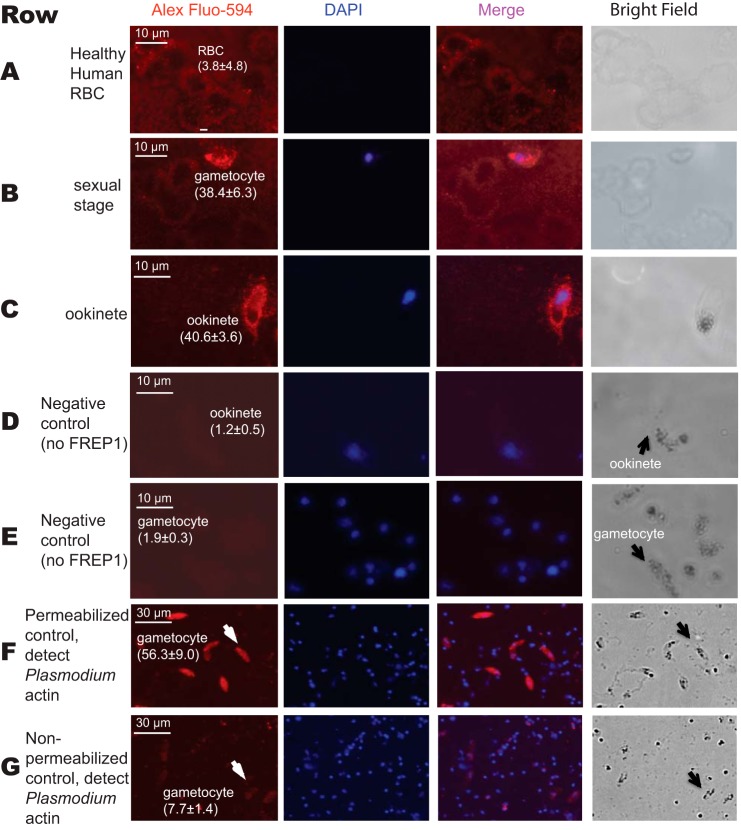
FIGURE 4.
Interaction between the recombinant FREP1 and P. falciparum demonstrated by immunofluorescence assays. Images in row A depict healthy, non-infected human RBC. Images in rows B and E depict sexual stage gametocytes. Images in the rows C and D are diploid ookinetes. Images in the rows D and E were obtained by conducting the same procedures as rows A–C except no recombinant FREP1 was added. Image F depicts the anti-actin antibody staining of gametocytes that were fixed with 100% methanol. Image G shows that anti-actin antibody does not stain malaria actin within gametocytes that were fixed with 4% paraformaldehyde. Of note, numerous DAPI-positive spots in rows F and G represent extracellular merozoite forms of P. falciparum, which become abundantly released after 10 days of culture. The first and second columns depict FREP1 and nuclear staining, respectively. Merging the first and second columns generated the third column. The last column shows cells under bright field. The numbers within parentheses after the cells are the average fluorescence signal intensity values (± S.D.) for three replicates.

