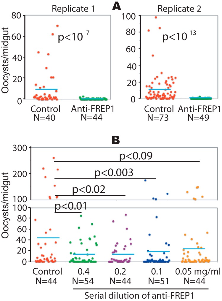
FIGURE 6.

The anti-FREP1 antibody blocks P. falciparum parasite invasion in mosquitoes. Three-day-old female An. gambiae were fed with P. falciparum-infected blood containing 0.2% stage V gametocytes and 0.5 mg/ml of purified anti-FREP1 antibodies (A) or a series of dilutions of anti-FREP1 antibodies (0.4, 0.2, 0.1, and 0.05 mg/ml) (B). Midguts from engorged (fed) mosquitoes were dissected 7 days post-infection and the numbers of oocysts in mosquito midguts were counted microscopically. N represents the number of mosquitoes and the blue bars represent the mean number of oocysts per midgut in each group. Control in panels A and B represent mosquitoes fed on infected blood treated with purified preimmune rabbit antibodies (0.5 and 0.4 mg/ml in A and B, respectively). The p values between experimental and the control groups were calculated using Wilcoxon-Mann-Whitney tests. Data represent two independent experiments.
