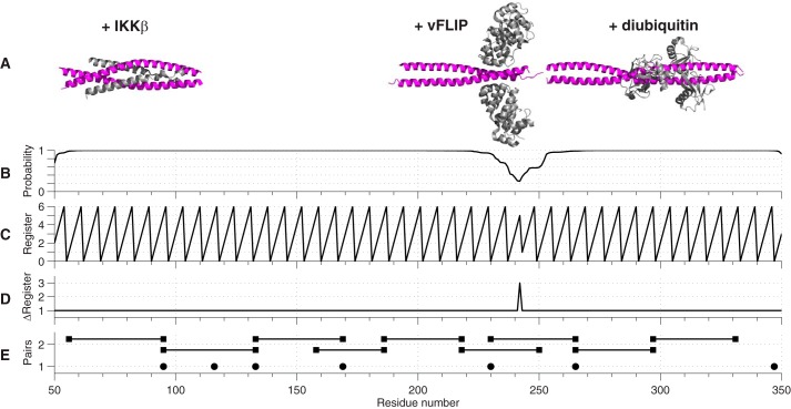
FIGURE 3.
A, crystal structures of fragments of IKKγ (magenta) in complex with IKKβ, vFLIP, and diubiuitin (gray) aligned according to their position in the sequence. B, probability of a region adopting a coiled-coil arrangement based on a window of 28 residues predicted from the primary sequence of IKKγ. C, predicted position of residues within a coiled-coil based on the heptameric repeat of the motif: each residue has a predicted position from 0–6 and a regular zigzag pattern implies a perfect coiled-coil. D, increment of the register of a residue with respect to its predecessor. An increment of +1 implies a perfect coiled-coil. E, positions of nitroxide spin labels for singly labeled IKKγ (solid circles) and doubly labeled IKKγ (solid squares joined by solid lines).

