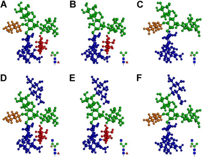
FIGURE 2.

Three-dimensional models do not show significant differences in the conformations of the N-glycans in the mutants and Col-0. A and D, predicted conformations of N-glycans with Man3XylFuc(GlcNAc)2 and GlcNAcMan3XylFuc(GlcNAc)2 structures, respectively, in Col-0. B and E, predicted conformations of N-glycans with Man3Fuc(GlcNAc)2 and GlcNAcMan3Fuc(GlcNAc)2 structures, respectively, in xylt-1. C and F, predicted conformations of N-glycans with Man3Xyl(GlcNAc)2 and GlcNAcMan3Xyl(GlcNAc)2 structures, respectively, in fab. GlcNAc, mannose, xylose, and fucose residues are indicated with dark blue, green, brown, and dark red, respectively.
