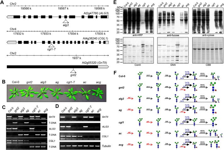
FIGURE 4.
Antibodies show different binding affinities for the proteins extracted from Col-0, gnt2, alg3, ag, cgl1, ac, and acg. A, schematic representation of the T-DNA insertion sites in the ALG3, CGL1 (GnTI), and GnTII genes. Boxes represent exons, and black denotes the coding region. T-DNA insertion sites and left border directions are indicated with triangles and arrowheads. B, phenotypes of the mutant plants compared with that of Col-0. Plants were grown on soil for 30 days. gnt2, alg3, and cgl-1T single mutants were used to produce ag, ac, and acg. C, PCR-based genotyping of the mutants. Gene-specific primers were designed and used in combination with T-DNA left border primer. D, RT-PCR analysis of the mutants. Levels of transcripts in Col-0 and the indicated mutants were determined by RT-PCR with gene-specific primers. Tubulin was used as a control. E, immunoblot and lectin blot analyses to assess structure and amount of the N-glycans in Col-0, gnt2, alg3, ag, cgl1, ac, and acg. Total proteins extracted from 3-week-old seedlings were subjected to immunoblot and lectin blot analyses. The immunoblots were probed with anti-HRP, anti-xylose, and anti-fucose antibodies, and lectin blots were probed with ConA and GNA. Coomassie Brilliant Blue (CBB) staining was used to show equal loading of the proteins. F, schematic illustration of the N-glycosylation in the mutant plants. N-Glycan processing in the indicated plants is illustrated with corresponding enzymes and structures of the N-glycans. The additions of the outer α1,3- and α1,6-mannose residues of the core α1,6-mannose of the N-glycan precursor are completely inhibited in alg3, addition of the 3-arm β1,2-GlcNAc residue of the N-glycan is completely inhibited in cgl1, and addition of the 6-arm β1,2-GlcNAc residue is completely inhibited in gnt2. The dominant additions of the core β1,2-xylose and α1,3-fucose residues are indicated with thick solid lines, and limited addition of the 6-arm β1,2-GlcNAc residue is indicated with thin dashed lines. In all mutants with the gnt2 background, the amount of PNGXF has been increased, indicating that the additions of the core β1,2-xylose and α1,3-fucose residues are regulated by the addition of the 6-arm β1,2-GlcNAc residue. Changed amounts of the N-glycans are represented by the enlarged and compressed N-glycan structures.

