
FIGURE 4.
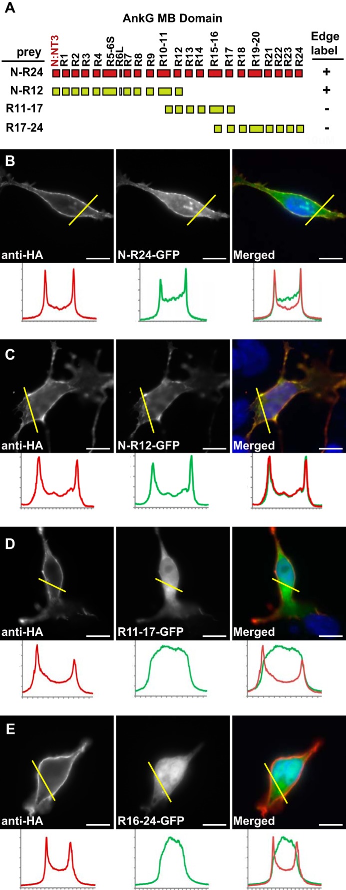
The N-terminal half of AnkG-MB (N-R12, ∼400 residues) contains the KCNQ2 determinant(s) responsible for surface redistribution. A, schematic showing MB domain exonic structure, encoded ankyrin repeats, and the overlapping truncations used to generate GFP fusion proteins. + indicates surface redistribution of GFP signal to the surface membrane was observed; − indicates GFP signal remained homogeneously cytoplasmic. B–E, surface redistribution assay results. The left panels show cells labeled using an extracellular HA tag on the neurofascin-KCNQ2 fusion protein bait, and middle panels show the distribution of AnkG-GFP fusion proteins, right panels show overlays (AnkG-GFP, green; anti-HA, red). Below each panel, intensity histograms show signals along the yellow line. B and C, AnkG-MB (green) and N-R12 truncation (green) are redistributed to the cell surface. D and E, MB R11–17 (green) and MB R16–24 (green) remain cytoplasmic. Scale bars, 10 μm.