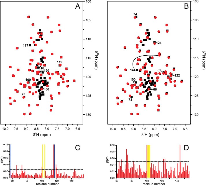
FIGURE 5.
Comparison of the Par14 and Par17 backbone amide resonances. In the two upper panels, the amide signal positions in the 1H,15N HSQC spectrum of Par17 (Biological Magnetic Resonance Bank (BMRB) accession code 18615; black squares) are overlaid with those of Par14 (red circles) based on the assignment by Terada et al. (14) (A) and Sekerina et al. (Ref. 17; Biological Magnetic Resonance Bank (BMRB) accession code 4768) (B). Shifted peaks are labeled with the respective Par17 residue number and in some cases connected by a line to denote the displacement. Signals belonging to the Par17 N terminus, which corresponds to the segment missing in Par14, are generally found as non-superposed black squares in the 1H resonance range between 8.5 and 8.0 ppm, thus indicating a random-coil structure. In the two lower panels, a CSP analysis for each residue within the structured PPIase domain again compares Par17 with the Par14 assignments of Terada (C) and Sekerina (D). Residues, whose backbone amide resonance assignments are missing in either Par17 or Par14 are indicated by yellow and blue bars, respectively.

