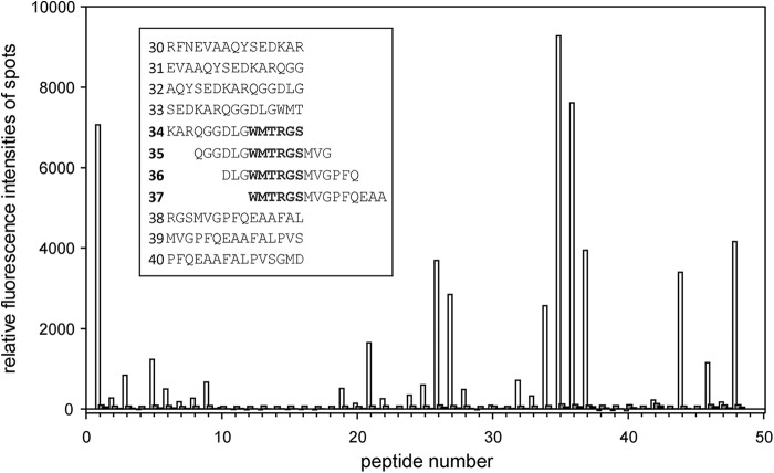
FIGURE 8.
Peptide microarray binding pattern of Ca2+/CaM. To display Ca2+/CaM binding to the microarray of Par17 15-mer peptides, relative fluorescence intensities of spots, which were corrected for the background, are plotted against the peptide number. Incubation of peptide microarrays were performed in the presence of either Ca2+ (white bars) or the Ca2+-chelator EGTA (black bars). As a control, the array was incubated only with anti-mouse-IgG (gray bars). The inset shows the amino acid sequences of peptides 30–40, comprising the dominant binding site for Ca2+/CaM in the Par17 peptide microarray.

