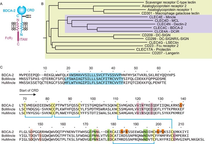
FIGURE 1.
Sequence of human BDCA-2. A, summary of the organization of BDCA-2 and other type 2 transmembrane receptors containing C-type CRDs. B, dendrogram showing the relationships between the CRD portions of this group of C-type lectins. The subgroup containing mincle and BDCA-2 is highlighted in the violet box. C, sequence of BDCA-2 compared with sequences of cow and human mincle. The N terminus of the CRD is denoted by the arrow. Disulfide bonds are indicated by blue lines between conserved cysteine residues, which are highlighted in yellow. Amino acid residues that create the conserved Ca2+-binding site are indicated in green, and residues in mincle that create the two accessory Ca2+-binding sites in mincle are highlighted in violet and pink. Residues that form part of the extended sugar-binding site of BDCA-2 are highlighted with orange. The putative transmembrane domains are shown in blue.

