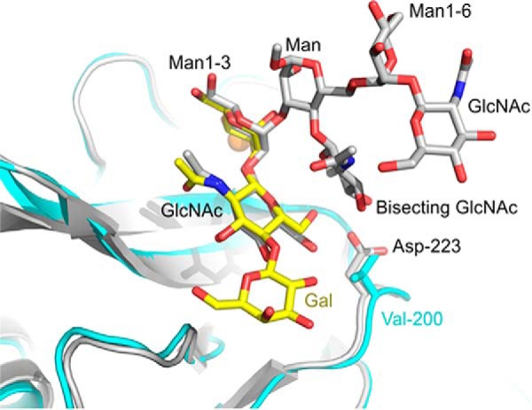
FIGURE 10.

Comparison of ligand binding sites in human BDCA-2 and mouse DCIR2. The structure of mouse DCIR2 is taken from Protein Data Bank entry 3VYK and is shown in gray. BDCA-2 is shown in cyan. In the superposed structures, carbon atoms of Galβ1–4GlcNAcβ1–2Man bound to BDCA-2 are in yellow, and carbon atoms of the biantennary glycan with bisecting GlcNAc, bound to DCIR2, are in gray. Ca2+ is shown in orange, oxygen atoms in red, and nitrogen atoms are dark blue.
