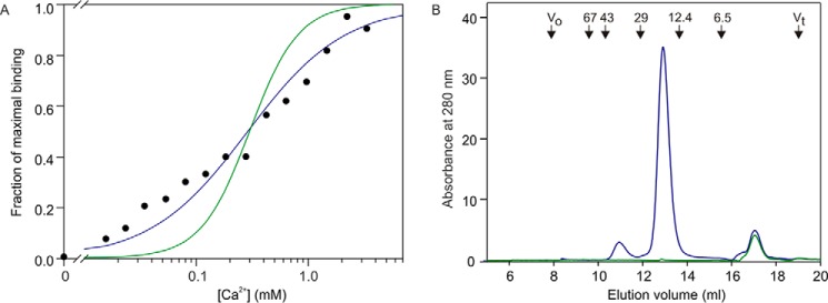
FIGURE 6.

Ca2+ dependence and gel filtration analysis of the CRD from BDCA-2. A, Ca2+ dependence of 125I-Man-BSA binding to BDCA-2. Binding of 125I-mannose-BSA to the biotin-tagged CRDs immobilized in streptavidin-coated wells was quantified. After binding of ligand in 150 mm NaCl and 25 mm Tris-Cl, pH 7.8, in the presence of various concentrations of Ca2+, wells were washed with buffer containing 150 mm NaCl, 25 mm Tris-Cl, pH 7.8, and 25 mm CaCl2. The experimental data, shown as black circles, were fitted to first and second order binding equations, shown respectively as blue and green lines, using a nonlinear least squares fitting program. B, gel filtration analysis of the CRD from BDCA-2 on a 7.5 × 300-mm column of Sephacryl S75 eluted with 100 mm NaCl, 10 mm Tris-Cl, pH 7.8, and 2.5 mm EDTA at a flow rate of 0.5 ml/min. Blue trace is with protein, and green trace is a mock sample without protein, showing that the peak eluting at 17 ml results from the presence of Ca2+-EDTA complex in the sample. Positions of molecular weight standards are shown at the top.
