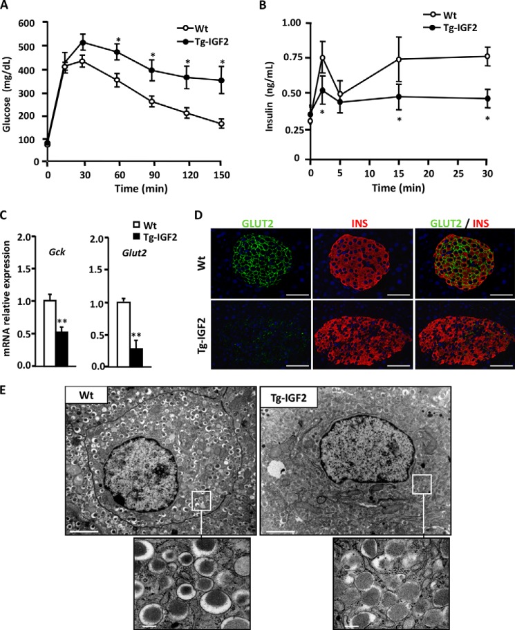
FIGURE 1.
Local overexpression of IGF2 alters islet functionality. A, Tg-IGF2 mice showed altered glucose tolerance test at 4 months of age. Three-month-old WT (white circle) and Tg-IGF2 (black circle) mice were given an intraperitoneal injection of 2 mg of glucose/g bw, and blood glucose was measured at the indicated times as described under “Experimental Procedures.” Results are expressed as mean ± S.E. of 10 animals/group. *, p < 0.05. B, in vivo insulin release test after intraperitoneal glucose injection (3 g/kg bw) to 4-month-old WT mice (white circles) and Tg-IGF2 mice (black circles), n = 10 animals/group (*, p < 0.05). C, analysis of the expression of β-cell glucose sensor genes, Gck and Slc2a2/Glut2, by qPCR on WT (white bars) and Tg-IGF2 (black bars) islets from 4-month-old mice. Data are mean ± S.E. of at least three pools of islets from eight animals/group. **, p < 0.01. D, immunohistochemical detection of INS (red) and GLUT2 (green) in pancreatic sections of 3-month-old WT and Tg-IGF2 mice. Original magnification ×400 (scale bar, 50 μm). E, electron microscopy of β-cells in pancreatic islets isolated from 4-month-old WT and Tg-IGF2 mice. Tg-IGF2 β-cells presented altered ultrastructure (×10,000) (scale bar, 2 μm). Inset images show secretory granules in more detail (×80,000) (scale bar, 0.2 μm).

