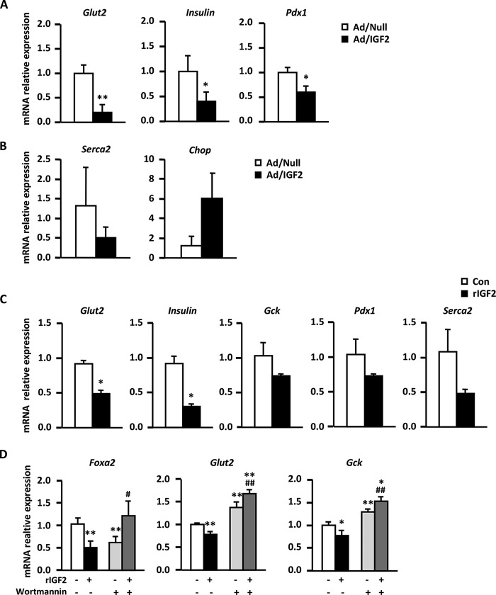
FIGURE 4.
IGF2 effects on adult WT islets. A, expression of β-cell markers by qPCR analysis in WT islets transduced with null control vector (Ad/Null, white bars) or adenovirus expressing IGF2 (Ad/IGF2, black bars). B, expression of ER stress genes by qPCR in the same adenovirus-transduced islets. Three pools of islets from eight animals/group were used. C, gene expression analysis of β-cell markers by qPCR in WT islets incubated for 48 h with either vehicle (white bars) or 100 ng/ml recombinant IGF2 protein (black bars). Three pools of islets from eight animals/group were used. D, analysis by qPCR of Foxa2, Slc2a2/Glut2, and Gck genes in INS-1 cells treated for 48 h with vehicle (white bars), 100 ng/ml recombinant IGF2 protein (black bars), 200 nm of the PI3K inhibitor wortmannin (light gray bars), or both (dark gray bars). Results shown represent the data obtained for at least six wells/condition and from three independent experiments. Data are expressed as mean ± S.E. *, p < 0.05; **, p < 0.01 versus vehicle-treated cells. #, p < 0.05, and ##, p <0.01 versus recombinant IGF2-treated cells.

