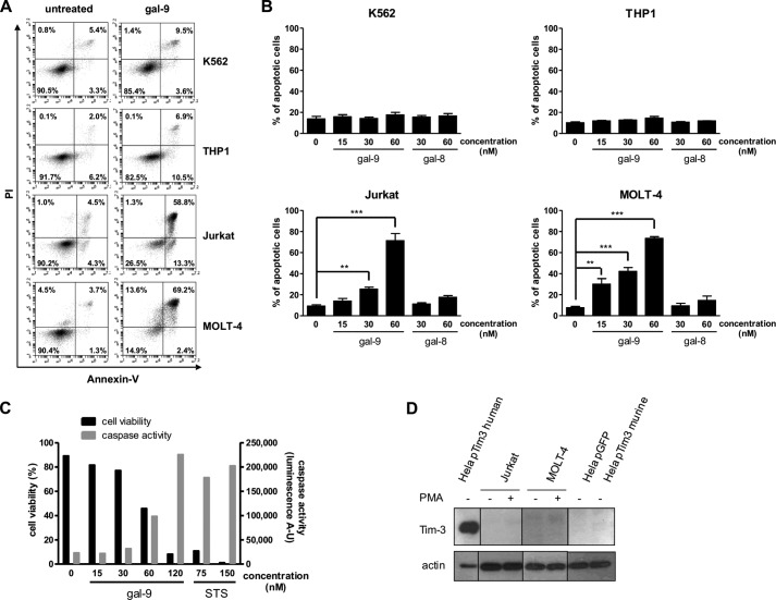
FIGURE 1.
Differential induction of apoptosis by gal-9 in lymphoid T cells and myeloid cells. A and B, two myeloid (K562 and THP1) and two lymphoid (Jurkat and MOLT-4) cell lines were treated with various amounts of recombinant gal-9 or gal-8 in serum-free medium for 24 h. Treated cells were stained with annexin-V/PI and then analyzed by flow cytometry. A, one example of flow cytometry plots for untreated cells (left panels) or cells treated with gal-9 (60 nm; right panels) following staining with annexin-V and PI. Bottom right quadrants are representative of early apoptotic cells (annexin-V-positive only) and top right quadrants of late apoptotic cells (annexin-V- and PI-positive). B, comparison of the percentages of apoptotic cells in the four lymphoid and myeloid cell lines treated with recombinant gal-9 or gal-8. Apoptotic cells were counted by flow cytometry as in A, taking into account all annexin-V-positive cells. Data are presented as means ± S.E. of three independent experiments. **, p < 0.01; ***, p < 0.001, one-way ANOVA followed by Dunnett's post hoc test. C, simultaneous measurement of cell viability and caspase-3/7 activity in Jurkat cells treated with increasing concentrations of gal-9 (0 to 120 nm) or staurosporine (STS; 75 or 150 nm). Following 24 h of treatment, cells were subjected to apoptosis measurement using two assays in parallel, annexin-V/PI staining and a caspase-3/7 enzymatic assay. The percentage of annexin-V-negative cells is represented on the left y axis (cell viability). Values of light emission (luminescence arbitrary units (A-U)) are plotted on the right y axis and are proportional to the level of activated caspase-3 and -7. The luminescence background value was measured using culture medium without cells. It was at 63 ± 5. D, Jurkat and MOLT-4 cells were treated or not with PMA for 24 h before Tim-3 expression analysis by Western blot. HeLa cells transiently transfected either with a control plasmid coding for GFP (pGFP) or a plasmid coding for human Tim-3 (pTim3 human) or murine Tim-3 (pTim3 murine) were used as positive and negative controls respectively, as the anti-Tim-3 antibody reacts against the human protein only. Actin protein levels were used as loading controls.

