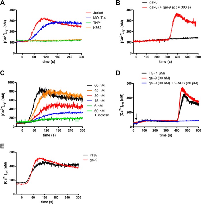
FIGURE 2.
Impact of gal-9 on ([Ca2+]cyt) in lymphoid T cells. A, variations of cytosolic calcium concentrations following gal-9 addition to lymphoid and myeloid cells (Jurkat/MOLT-4 and THP1/K562, respectively). [Ca2+]cyt was assessed by Indo-1 fluorimetry in the presence of 1 mm extracellular CaCl2. Gal-9 (30 nm final) was added at t = 0 s. B, variations of [Ca2+]cyt in Jurkat cells treated with gal-8 (30 nm, added at t = 0 s) alone (black line) or with gal-8 combined with secondary addition of gal-9 (30 nm, added at t = 300 s) (red line). C, dose/effects relationships in Jurkat cells treated with increasing concentrations (6–60 nm) of gal-9. [Ca2+]cyt was recorded in the presence of 1 mm extracellular CaCl2. The rise in [Ca2+]cyt was totally abolished by preincubation of gal-9 (30 nm) with a saturating concentration of lactose (5 mm). D, comparison of the impact of thapsigargin and gal-9 on calcium mobilization in Jurkat cells. Treatment of Jurkat cells with TG (1 μm) or gal-9 (30 nm) with or without 2-aminoethoxydiphenyl borate (2-APB) (30 μm) and in the absence of extracellular CaCl2 is indicated with a black arrow. Re-addition of CaCl2 was performed at t = 400 s. E, comparison of the impact of PHA and gal-9 on calcium mobilization in Jurkat cells. Variations of [Ca2+]cyt in Jurkat cells were treated with gal-9 (30 nm) or PHA (10 μg/ml) in the presence of 1 mm extracellular CaCl2. All the experiments depicted in this figure are representative of at least two similar independent experiments.

