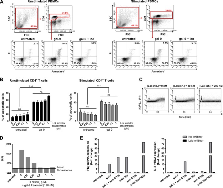
FIGURE 7.
Contribution of Lck to peripheral CD4+ T cell response to gal-9. A and B, PBMCs freshly isolated from blood were stimulated or not with anti-CD3 and anti-CD28 antibodies for 48 h. A, resting or activated PBMCs were preincubated or not with lactose (5 mm) prior to gal-9 treatment (30 nm) for 24 h. The percentage of apoptotic cells was determined by flow cytometry (annexin-V/PI staining) after gating on the CD4+ population. Flow cytometry plots are representative of four independent experiments realized with different healthy donors. B, impact of the Lck inhibitor on gal-9-induced apoptosis in CD4+ T cells. Resting or activated PBMCs were preincubated or not with the Lck inhibitor (0.01 to 1 μm) for 1 h before addition of gal-9 (30 nm) and incubation for 24 h. The percentages of apoptotic cells were determined as in A, taking account of all annexin-V-positive cells. Histograms represent means ± S.E. of three independent experiments with different donors. Statistical analyses using two-way ANOVA revealed no impact of the Lck inhibitor on the rate of apoptosis within the gal-9-untreated or gal-9-treated groups and a strong impact of gal-9 regardless of the presence or absence of the Lck inhibitor. ***, p < 0.001. ns, not significant. C and D, gal-9 induced calcium mobilization in peripheral CD4+ T cells, which was blocked by Lck inhibition. Resting PBMCs were preincubated with Fluo4-AM (1 μm) for 45 min at room temperature. After washing and CD4 staining, cells were treated or not with the Lck inhibitor (0–2 μm) for 30 min before starting gal-9 stimulation (120 nm). C, fluorescence intensity (F-I; proportional to [Ca2+]cyt) continuous recording by flow cytometry after gal-9 addition (arrow). Plots are representative of [Ca2+]cyt variations in CD4+ T cells in two independent experiments. D, mean fluorescence intensity (MFI) recorded in CD4+ T cells measured on the different plots as in C, with the flow cytometer software. E, gal-9 induction of IL-2 and IFNγ transcription and the impact of the Lck inhibitor. CD4+ T cells isolated from resting PBMCs were preincubated or not with the Lck inhibitor (500 nm) for 1 h before treatment with gal-9 (30 nm) or the combination of anti-CD3 and anti-CD28 antibodies (1 μg/ml) for 6 h. IL-2 and IFNγ mRNA expression were determined by quantitative real time PCR. Data presented here correspond to the fold change defined as the ratio of target gene mRNA with that of untreated cells (the housekeeping gene TBP was used as an internal control). These results are representative of two independent experiments.

