FIGURE 7.
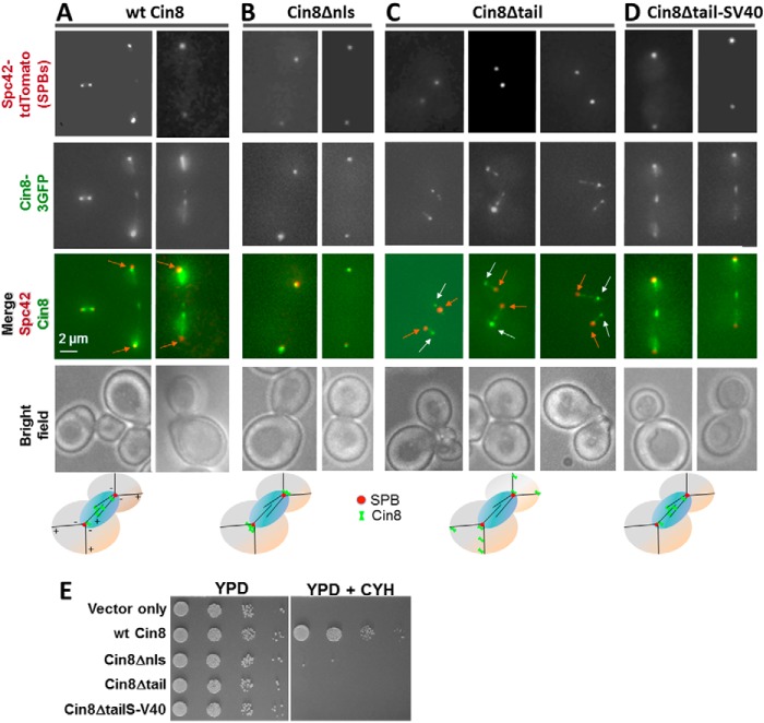
In vivo localization and viability of Cin8 variants. A–D, deletion of the tail of Cin8 shifts its localization to the plus-ends of the MTs. Representative two-dimensional projections of cells co-expressing GFP-tagged variants of wt Cin8 and the SPB-localizing Spc42-tdTomato are shown. Cin8 variants are indicated on the top of each panel (see Fig. 1B for description). A, wt Cin8. B, Cin8Δnls. C, Cin8Δtail. D, Cin8Δtail-SV40. Orange arrows point toward the SPBs; white arrows point to the accumulation of Cin8 at the plus-ends of cytoplasmic MTs. Bottom row: model for intracellular localization of the different variants of Cin8. E, viability of cin8Δkip1Δ cells expressing vector only or 3GFP-tagged variants of Cin8 from a CEN plasmid. Cells were grown in serial dilutions on either YPD plates (left) or YPD plates containing 7.5 μg/ml cycloheximide (CYH, right).

