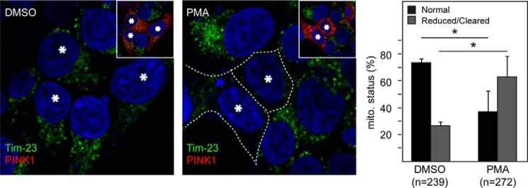
FIGURE 8.

Pharmacological NF-κB activation promotes mitochondrial clearance in PINK1-expressing cells. Representative confocal images of HEK293 cells transiently transfected with untagged PINK1 treated with DMSO or 10 ng/ml PMA for 24 h (PINK1-transfected cells are denoted by asterisks and revealed by anti-PINK1 staining shown in the inset). The outline indicates the reduced mitochondrial network in PINK1-transfected cells treated with PMA. The percentage of cells showing normal or reduced/cleared mitochondria is shown in the right panel. The total number of cells analyzed (n) is indicated. This experiment was repeated three times (*, p < 0.05).
