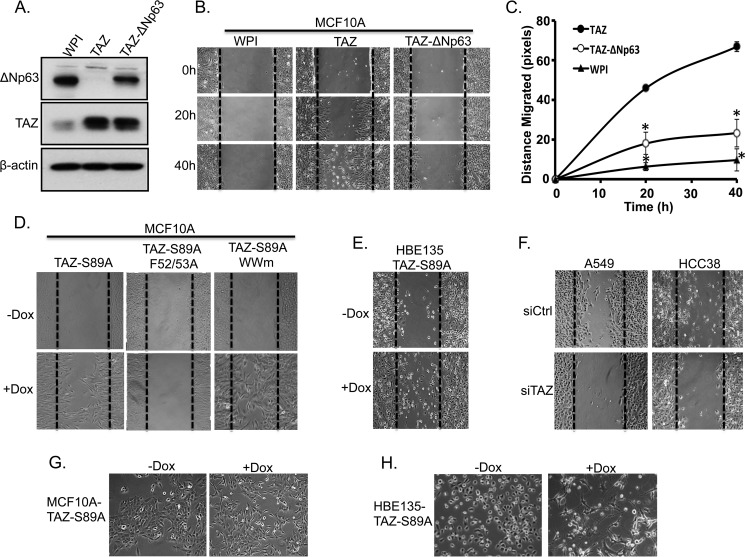
FIGURE 7.
Reintroduction of ΔNp63 partially recues TAZ-mediated cell migration. A, Western blot analysis of ΔNp63 expression. MCF10A-TAZ cells were infected with lentivirus expressing ΔNp63 (MCF10A-TAZ-ΔNp63). Protein was extracted from these cells, and ΔNp63 expression was compared with MCF10A-WPI and MCF10A-TAZ cells. β-Actin was used as an internal loading control. B, ΔNp63 reintroduction in TAZ-overexpressing cells partially rescues TAZ-induced increased cell migration. MCF10A-WPI, MCF10A-TAZ, and MCF10A-TAZ-ΔNp63 cells were plated to confluence and starved in 2% horse serum overnight. A wound healing assay was performed, and cell migration was analyzed between cells at different time points (0, 20, and 40 h). C, quantification of cell migration. Cell migration distance (pixels) was quantified in all cells as described in B. The experiment was performed in triplicate, and error bars represent S.D. from each set of triplicates. *, statistically significant difference (p < 0.05) between MCF10A-TAZ and MCF10A-WPI or MCF10A-TAZ-ΔNp63. D, TEAD-dependent increased cell migration by TAZ. Cell migration analyses were performed using the established cell lines and conditions described in Fig. 3A. E, overexpression of TAZ-S89A causes increased cell migration in HBE135 cells. Wound healing analyses were performed in cell lines described in Fig. 2C. F, knockdown of TAZ in HCC38 and A549 cells decreases cell migration. G and H, overexpression of TAZ-S89A induces EMT in both MCF10A (G) and HBE135 (H) cells.

