
FIGURE 1.
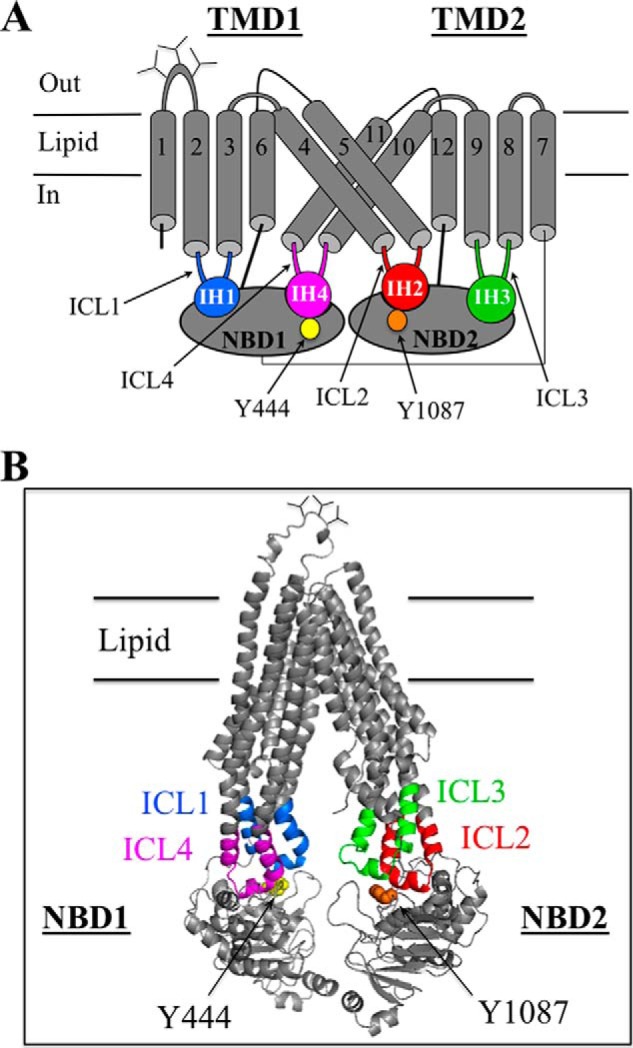
Models of human P-gp. A, secondary structure of human P-gp showing the four ball-and-socket joints of the NBD-TMD transmission interfaces. The intracellular loops (ICLs) containing intracellular helices (IHs) that interact with the NBDs are shown in color. The cylinders represent TM segments, and the branched lines in the loops connecting TM segments 1 and 2 represent glycosylated sites. B, predicted structure of human P-gp in an open conformation was based on the crystal structure of mouse P-gp (56). The intracellular loops interacting with the NBDs are colored. The model was viewed using the PyMol system (57).