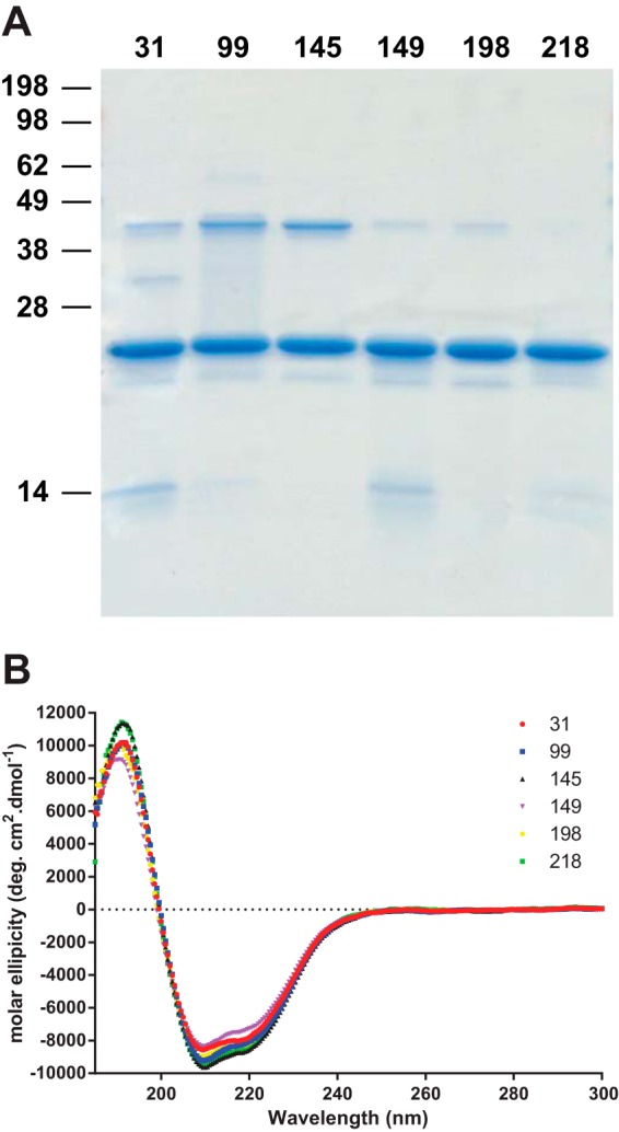
FIGURE 2.

Characterization of the NBD-labeled proteins. A, Coomassie Blue-stained 16% polyacrylamide Tris-glycine gel showing NBD-labeled protein. Prior to loading, samples were heated to 95 °C for 5 min in SDS loading buffer in the absence of reducing agent. B, circular dichroism spectra of the PrP proteins labeled at the different sites. deg, degrees.
