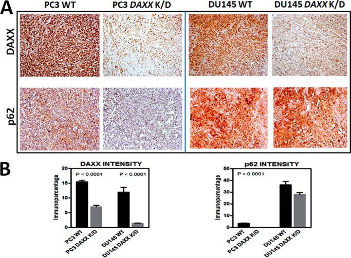
FIGURE 3.
DAXX modulates autophagy markers in vivo. A, quantitative immunohistochemical analysis of protein markers was performed using excised tumor tissue corresponding to WT (n = 6) and DAXX K/D (n = 6) tumors from either PC3 or DU145 injections. Tissue sections were stained using antibodies specific for DAXX or p62. Representative ×10 images are shown. B, immunostaining was quantified and analyzed using Aperio ImageScope software. To construct graphs, GraphPad Prism version 5 software was used. Statistical significance was assessed by unpaired Student's t test. Note that DU145 cells, being autophagy-defective, show high levels of p62, regardless of the DAXX status. Error bars, S.E.

