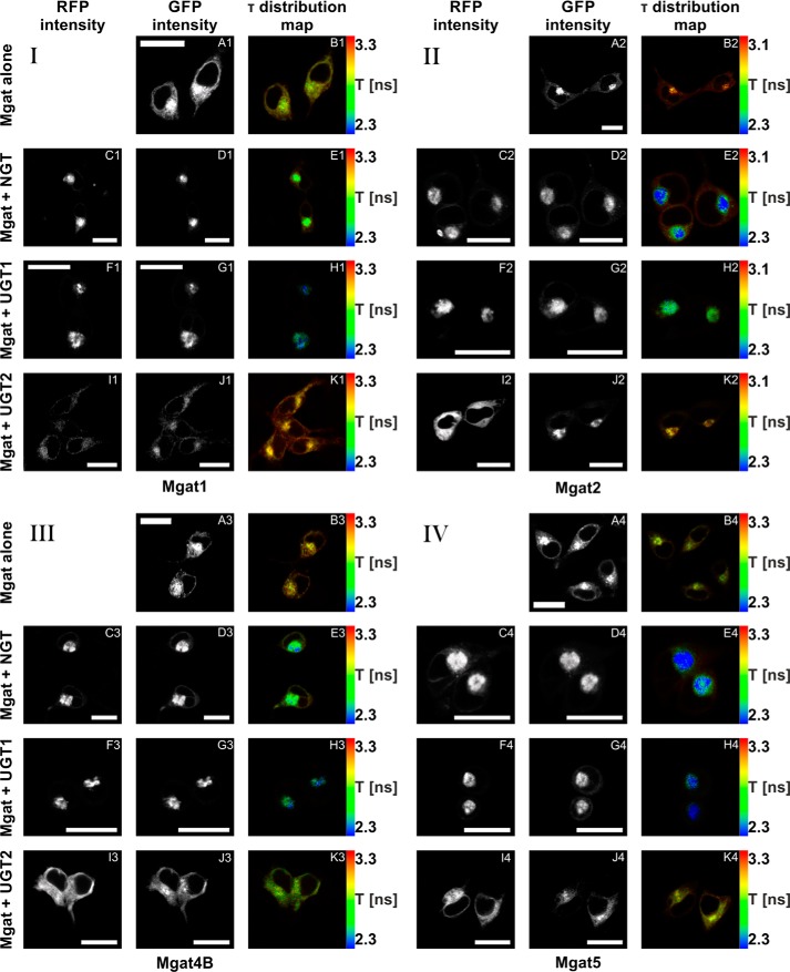
FIGURE 4.
In vivo FLIM-FRET analysis of interactions between Mgats and UDP-sugar transporters. A–K, confocal intensity-resolved (A, C, D, F, G, I, and J) and time-resolved (B, E, H, and K) imaging of eGFP-Mgat (D, G, and J) interaction with mRFP-UDP-sugar transporter (C, F, and I) in HEK293T cells in comparison with cells expressing eGFP-Mgat only (A). FLIM-FRET analysis of either Mgat1 (panel I), Mgat2 (panel II), Mgat4B (panel III), or Mgat5 (panel IV) putative interactions with NGT (C–E), UGT1 (F–H), and UGT2 (I–K). GFP fluorescence lifetime (τ) was shortened by simultaneous overexpression of eGFP-Mgats with both mRFP-NGT and mRFP-UGT1, strongly suggesting an interaction between these proteins. In the case of mRFP-UGT2, the same phenomenon was demonstrated only upon eGFP-Mgat4B co-expression. Co-expression of mRFP-UGT2 with eGFP-Mgat1, Mgat2, and Mgat5 did not influence GFP fluorescence lifetime. The red to blue color shift reflects shortening of the fluorescence lifetime. The rainbow scale bars placed next to time-resolved images (B, E, H, and K) represent fluorescence lifetime range between either 2.3 (blue) and 3.3 ns (red; Mgat1, Mgat4B, or Mgat5) or 2.3 (blue) and 3.1 ns (red; Mgat2). Bars, 20 μm. τ, fluorescence lifetime.

