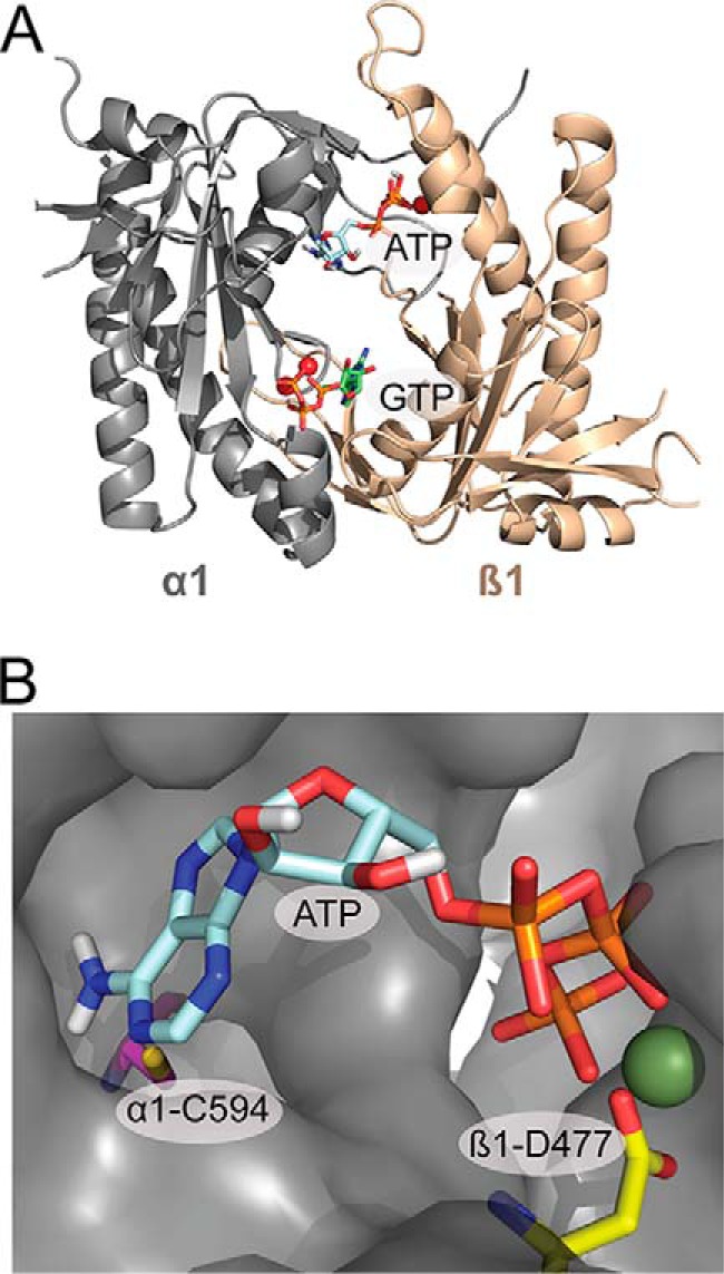
FIGURE 1.

Model of the catalytic domain of sGC in the active confirmation based on the crystal structure of mammalian adenylate cyclase (21). A, Model of the catalytic domain with ATP docked at the pseudosymmetric site, and GTP docked at the catalytic site into the model using Autodock 4 (58). B, ATP docked in the pseudosymmetric site cavity. The residues mutated in the pseudosymmetric site, α1-Cys-549 (magenta) and β1-Asp-477 (yellow), and the Mg2+ ion (green) are highlighted.
