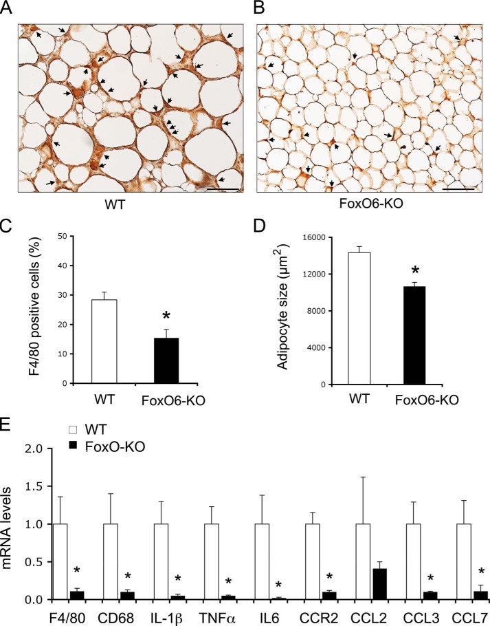
FIGURE 7.
Effect of FoxO6 depletion on macrophage infiltration in adipose tissues. Adipose tissues were procured from high fat-fed WT control (A) and FoxO6-KO mice (B) as described in Fig. 5. Paraffin-embedded adipose tissues were subjected to anti-F4/80 immunohistochemistry. F4/80-positive cells (stained brown) were scored and normalized to the total number of adipose cells (C). In addition, the size of adipose cells was determined and compared between control and FoxO6-KO groups (D). Epididymal adipose tissues were minced for the preparation of stromal vascular cells. Total RNA was isolated from stromal vascular cells and subjected to real-time qRT-PCR using 18S RNA as control for determining F4/80, CD68, IL-1β, TNFα, IL-6, CCR2, CCL2, CCL3, and CCL7 mRNA levels (E). *, p < 0.05 versus WT control. n = 8–9 per group. Bar, 100 μm.

