FIGURE 3.

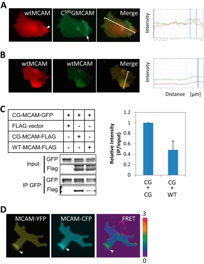
MCAM depalmitoylation promotes MCAM-MCAM interaction. A, wild type MCAM-YFP (red, arrowhead) localizes separately from palmitoylation-resistant C590G MCAM-CFP (green, arrow) in Wnt5a-treated melanoma cells. White lines, regions of interest used for the line scans. B, wild type MCAM-YFP (red) and wild type MCAM-CFP (green) co-localize asymmetrically in Wnt5a-treated WM239A melanoma cells. White lines, regions of interest used for the line scans. C, C590G MCAM-GFP forms a complex with C590G MCAM-FLAG with higher affinity than with wild type MCAM-FLAG in HEK293T cells (Student's t test, p = 0.006, average of three experiments). D, interaction between MCAM molecules detected by live CFP/YFP FRET imaging of melanoma cells expressing MCAM-YFP and MCAM-CFP. Increased FRET signal was detected at the area of asymmetrically localized MCAM (arrows). IP, immunoprecipitation. Error bars, S.E.
