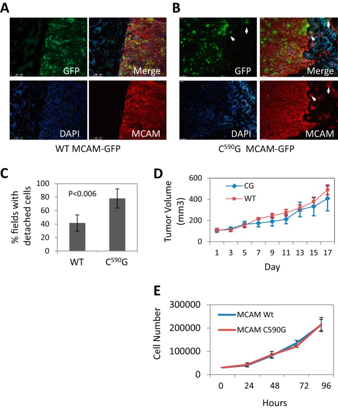
FIGURE 5.

Blocking MCAM palmitoylation promotes melanoma invasion in vivo. A, xenograft tumors stained for endogenous MCAM (red) and WT MCAM-GFP (green). B, C590G MCAM-GFP-expressing tumors have irregular borders with C590G MCAM-GFP-expressing cells (green) detached (arrows) from the primary tumor residing in the adjacent mouse tissue (DAPI, blue). C, quantitation of detached tumor cells (average fields/tumor). Error bars, S.D. p value determined by Student's t test. D, xenograft tumor volumes, measured when first palpable (day 1). Error bars, S.D. E, growth rate of WT and C590G MCAM-GFP-expressing cells in two-dimensional culture.
