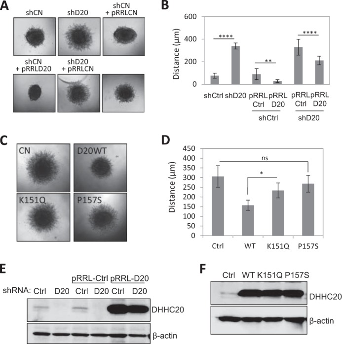
FIGURE 7.

DHHC20 inhibits cell invasion. A, inhibition of DHHC20 increases cell invasion in three-dimensional collagen compared with control shRNA. Spheroid invasion was quantified 8 days following transfer into collagen. B, quantification of spheroids in A. Distance was measured from the edge of the core to the invasion front (shCtrl (n = 10), shDHHC20 (n = 11), shCtrl + pRRLCtrl (n = 12), shCtrl + pRRLD20 (n = 12), shDHHC20 + pRRLCtrl (n = 9), and shDHHC20 + pRRLD20 (n = 8)). Statistical analysis was performed using a one-way ANOVA with Tukey's multiple-comparison test (**, p < 0.01; ****, p < 0.0001). pRRLCtrl is a lentiviral vector expressing GFP, and pRRLD20 is a lentiviral vector expressing DHHC20 that is resistant to shRNA-mediated silencing. C, spheroid invasion assays of C590G MCAM-GFP-expressing cells expressing control pRRL-vector alone (Ctrl), WT, K151Q, or P157S mutant DHHC20 (D20). D, quantification of spheroids in C (*, p = 0.012; ns, p = 0.369). p values were determined using one-way ANOVA with Tukey's multiple-comparison test. Error bars, S.D. E, Western blot showing expression levels of DHHC20 in the cell lines used in A. F, Western blot showing expression levels of WT DHHC20 and the mutant forms of DHHC20 in the cell lines used in C.
