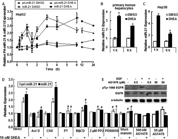
FIGURE 1.
DHEA increases pri-miR-21 transcription and mature miR-21. For all panels, cells were serum-starved, i.e. incubated in phenol-red free DMEM medium containing 5% dextran charcoal-stripped FBS, for 48 h before treatment to reduce steroid hormone levels. qPCR was used to examine miR-21 and pri-miR-21 expression relative to RNU48 and 18S rRNA, respectively. A, HepG2 cells were incubated with DMSO (vehicle control) or 10 nm DHEA for the indicated times. Relative expression is -fold change normalized to DMSO at time zero. Values are the average of 2–4 separate experiments ± S.E. B, primary human hepatocytes (B) or Hep3B human hepatoma cells (C) were treated with DMSO or 10 nm DHEA for 1 or 6 h, and qPCR was performed for miR-21. Values are the average of three determinations within one experiment ± S.E. *, p < 0.05 versus DMSO. D, where indicated, HepG2 cells were preincubated with the following inhibitors: 10 μg/ml ActD, 10 μg/ml CHX, 100 ng/ml PT, 10 μm MβCD, 500 nm AG-1478 (tyrphostin), 2 μm PP2, 50 μm PD98059, or 50 nm wortmannin before 1 h of treatment with DMSO or 10 nm DHEA. Values are the mean ± S.E. of three-four separate experiments/time point. *, p < 0.05 versus DMSO vehicle; #, p < 0.05 versus 10 nm DHEA. E, to confirm that AG1478 blocks EGFR activity in HepG2 cells at the concentrations used, cells were treated with 10 ng/ml EGF ± the indicated concentrations of AG1478 for 15 min. Whole cell lysates, 40 μg of protein, were immunoblotted first for Tyr(P)-1068-EGFR. The membrane was stripped and re-probed for EGFR.

