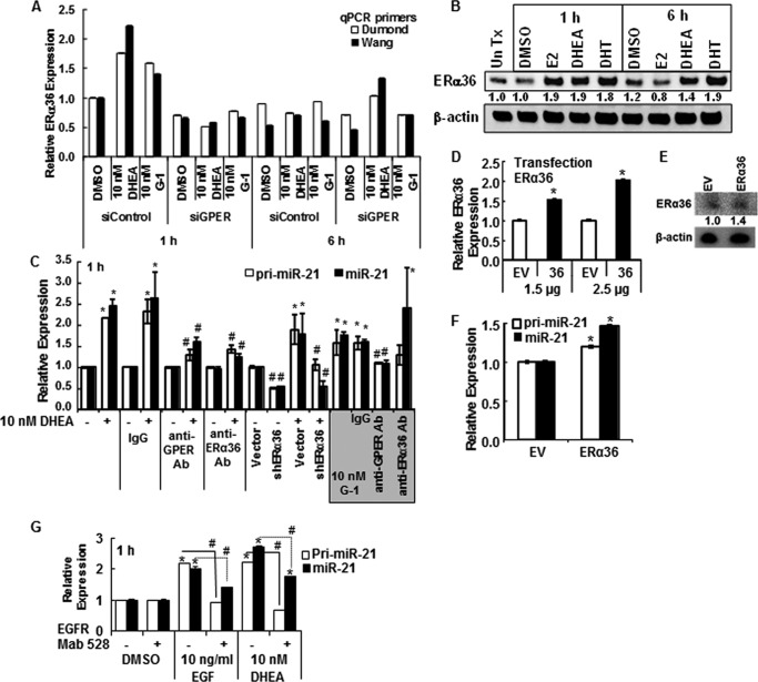
FIGURE 4.
DHEA activates PM-bound GPER and increases pri-miR-21 transcription. For all panels, HepG2 cells were serum-starved for 48 h before treatment. A, where indicated, HepG2 cells were transfected with siRNA control or siRNA against GPER for 48 h. Cells were treated with DMSO (vehicle control), 10 nm DHEA, or 10 nm G-1 for 1 or 6 h. qPCR used two different primer sets specific for ERα36 (Dumond (40) and Wang (41); see “Experimental Procedures”. Values are the average ± S.E. of triplicate determinations. B, Western blot for ERα36 using 25 μg of WCE from HepG2 cells treated with DMSO, 10 nm E2, 10 nm DHEA, or 10 nm DHT for 1 or 6 h. The membrane was stripped and re-probed for β-actin. The values are the ratio of ERα36/β-actin normalized to 1 h DMSO. C, HepG2 cells were preincubated with 1:200 dilution of rabbit IgG or antibodies (Ab) against GPER or ERα36 for 24 h and then treated with DMSO, 10 nm DHEA, or 10 nm G-1 (shaded gray) for 1 h. Where indicated cells were transfected with pRNAT-U6.1 vector control or the shERα36 plasmid for 48 h before 1 h treatment with DMSO or 10 nm DHEA. Values are the mean ± S.E. of three separate experiments. *, p < 0.05 versus DMSO control. #, p < 0.05 from the indicated treatment (DHEA or G-1) without antibody or control plasmid. D, HepG2 cells were transfected with the indicated amounts of empty expression vector (EV) or pCR3.1-ERα36 (37) for 48 h before qPCR for ERα36 relative to 18S. Values are the average ± S.E. of triplicate determinations. *, p < 0.05 versus empty vector control. E, Western blot of ERα36 relative to β-actin. F, transfection of HepG2 cells with ERα36 for 48 h increased pri-miR-21 and miR-21 levels. Values are the average ± S.E. of triplicate determinations. *, p < 0.05 versus empty vector control. G, EGF stimulation of pri-miR-21 and miR-21 expression is blocked by EGFR monoclonal antibody 528. Cells were incubated with 1 μg/ml EGFR antibody 528 or mouse IgG for 3 h before the addition of DMSO (vehicle control) or 10 nm DHEA for 1 h. Values are the average ± S.E. of triplicate determinations within one experiment. *, p < 0.05 versus DMSO vehicle; #, p < 0.05 versus EGF or DHEA, as indicated.

