FIGURE 7.
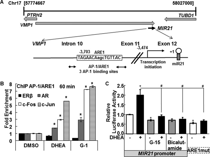
DHEA rapidly increases AR, c-Fos, and c-Jun recruitment to and dismisses ERβ from the miR-21 promoter. A, the diagram shows the chromosomal location of the pri-miR-21 promoter within the VMP2/TMEM49 gene (see the Atlas of Genetics and Cytogenetics in Oncology and Haematology). The location and sequences of the AP-1/ARE and the primers (black arrows) used for ChIP are indicated. MatInspector identified three AP-1 binding sites in the ARE region. B, HepG2 cells were serum-starved for 48 h and then treated with DMSO, 10 nm DHEA, or 10 nm G-1 for 60 min. ChIP was performed using antibodies for ERβ, AR, c-Fos, and c-Jun as described under “Experimental Procedures.” Values are -fold enrichment of the PCR product in the immunoprecipitated samples relative to input control and are the average ± S.E. of three separate experiments, performed in triplicate within each biological replicate. *, p < 0.05 versus DMSO. C, HepG2 cells were transfected with a firefly luciferase reporter constructs driven by the 5′-flanking region of human MIR21 in the sense orientation or with mutations in ARE1 (ARE1mut) (26). The cells were treated with DMSO (−, vehicle control) or 10 nm DHEA for 24 h. Where indicated, cells were preincubated for 15 min with 1 μm G-15 or 6 h with 100 nm bicalutamide (AR antagonist). The results are expressed relative to DMSO in the full-length MIR21 reporter after normalization with Renilla luciferase (mean ± S.E., n = triplicate wells in one experiment). *, p < 0.05 versus DMSO; #, p < 0.05 versus 10 nm DHEA.

