
FIGURE 1.
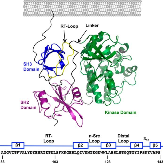
A, domain structure of inactive c-Src (PDB 2SRC). From N- to C terminus: SH3 domain (blue); SH2 domain (purple); kinase domain (green). The linker between the SH2 and kinase domains is colored black, and the RT-loop in the SH3 domain is colored yellow. The amino acid sequence of the human c-Src SH3 domain is numbered according to UniProtKB/Swiss-Prot entry P12931 and indicates the location of its five β strands and the interconnecting loops.