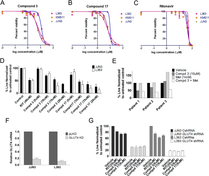
FIGURE 4.
Compounds 3 and 17 are more cytotoxic than ritonavir in multiple myeloma. The IC50 value of compound 3 (A), compound 17 (B), and ritonavir (C) was determined in L363, JJN3, and KMS11 cells by treatment with 0.39–50 μm compounds 3 or 17 or 1.25–100 μm ritonavir. Cell survival was examined after 72 h by Cell Titer-Glo luminescent cell viability assay. D, L363, JJN3, and KMS11 cells were cultured in the presence or absence of either ritonavir (40 μm), compound 3, or compound 17 (5, 10, 15, and 20 μm) in RPMI 1640 media containing 11 mm glucose and 2 mm glutamine for 72 h followed by assessment of viability by annexinV/DAPI staining. E, viability of CD38+ CD45− myeloma cells after a 48-h treatment with compound (Cmpd) 3 (10 μm) or metformin (Met) (1 mm) was assessed by annexinV DAPI staining. F, JJN3 and L363 cells were transduced with nontargeting or GLUT4-directed shRNA. Efficiency of KD was determined by quantitative PCR analysis of GLUT4 expression normalized to GAPDH expression. G, cells from F were utilized to determine impact on growth subsequent to indicated treatments after 72 h evaluated using trypan blue staining and an automated cell counter. Data, normalized to vehicle controls, represent the mean ± S.E. of three independent measurements.

