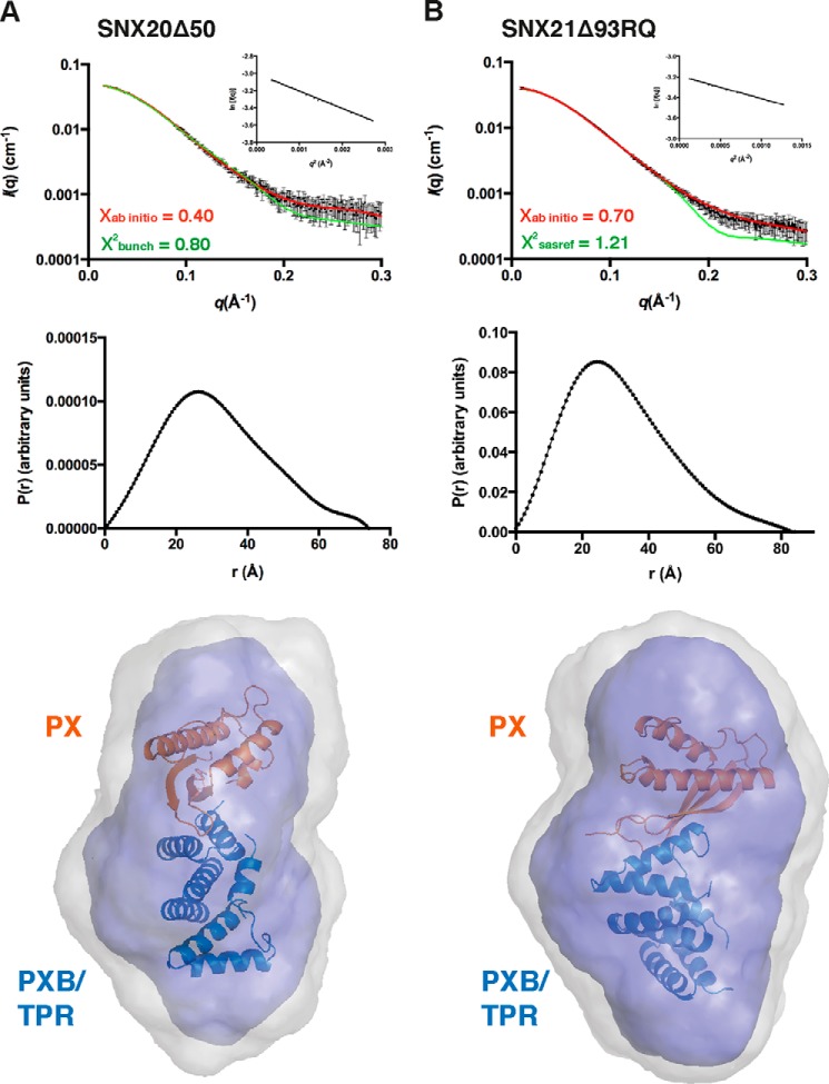
FIGURE 5.
SAXS solution structures of the SNX-PXB proteins. SAXS data for the SNX20Δ50 (A) and SNX21Δ93RQ (B) proteins. Top, experimental SAXS data (black) overlaid with the theoretical profiles calculated from rigid body modeling by CRYSOL (green), or the ab initio model determined by the program GASBOR (red). Middle, distance distribution functions P(r) calculated from the scattering data of SNX20Δ50 and SNX21Δ93RQ. Bottom, molecular models of the SNX-PXB proteins show their globular compact arrangement in solution. The SNX21 TPR domain crystal structure (blue) was combined with a model of the SNX20 PX domain (generated by Swiss-model, orange) using BUNCH, and overlaid with the ab initio structures of SNX20Δ50 and SNX21Δ93RQ.

