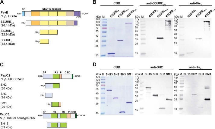
FIGURE 2.
Heterologously expressed PavB and PspC fragments. A and C, schematic model of PavB of S. pneumoniae TIGR4 and PspC subtypes PspC2 (S. pneumoniae ATCC33400) and PspC3 (S. pneumoniae D39 or serotype 35A) and heterologously expressed His6-tagged fragments of both surface proteins. SP, signal peptide; SSURE, streptococcal surface repeats; LPNTG, sortase anchoring motif; R, repeat domain; P, proline-rich sequence; CBD, choline-binding domain. B, SDS-PAGE of heterologously expressed PavB fragments (SSURE2, SSURE2+3, SSURE1–5) stained with CBB R250 and corresponding immunoblots. Proteins were detected using a specific polyclonal mouse anti-SSURE2+3 antibody or a monoclonal mouse anti-penta-His antibody and an alkaline phosphatase-coupled secondary anti-mouse antibody. D, SDS-PAGE of heterologously expressed PspC2 (SH2, SH3, SM1) and PspC3 (SH13) fragments stained with CBB R250 and corresponding immunoblots. Proteins were detected using a specific polyclonal mouse anti-SH2 antibody or a monoclonal mouse anti-penta-His antibody and an alkaline phosphatase-coupled secondary anti-mouse antibody.

