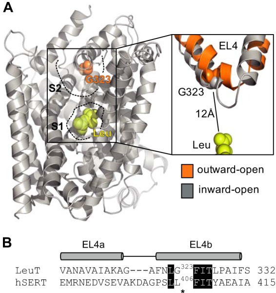
FIGURE 1.

Location of the L406E mutation. A, substrate-bound x-ray crystal structure of LeuT (PDB code 2A65). The substrate (leucine) binds in the central S1 site and is shown as yellow spheres. Gly-323 (which is equivalent to Leu-406 in human SERT) is located close to the tip of EL4 in the vestibular S2 site and is shown as orange spheres. An overlay of x-ray crystal structures of LeuT in outward-facing (shown in orange; PDB code 3TT1) and inward-facing (shown in gray; PDB code 3TT3) conformations is shown on the right to illustrate the flexibility of EL4. Gly-323 is located ∼12 Å away from the central substrate binding site. B, amino acid sequence alignment of the EL4 region in LeuT and human SERT (48). Identical residues are shown as white text on black background. The segments that assume α-helical arrangement in the LeuT x-ray crystal structures (denoted EL4a and EL4b) are indicated above the sequence alignment. Asterisks indicate the position of the Leu-406 residue (SERT numbering).
