FIGURE 4.
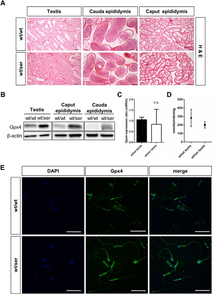
Immunohistological analysis of germinal epithelium and isolated spermatozoa. A, H&E analysis of testis and epididymis did not show any overt differences between wild type and homozygous tissues (scale bar = 100 μm). B, as observed for somatic tissues, Gpx4 immunoblotting in testis and epididymal tissue presented a stronger Gpx4 signal in heterozygous samples compared with wild type tissue. C, quantitative RT-PCR of Gpx4 levels in testicular tissue did not show a statistically significant difference between wild type and heterozygous testis. n.s., not significant. D, measurement of Gpx4-specific activity revealed a tendency toward decreased activity, but it did not reach statistical significance in heterozygous testicular tissue. E, whole mounts staining of epididymal spermatozoa revealed an increase in Gpx4 immunoreactivity (green) in heterozygous sperm that was mainly confined to the midpiece and the head region of sperm. Sperm nuclei were additionally counterstained with DAPI (scale bar = 50 μm).

