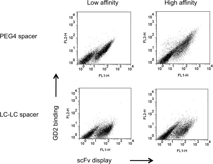
FIGURE 1.
Evidence of improved biotinylated GD2 with flexible PEG spacer. Yeast cells displayed low- and high-affinity hu3F8 scFv with c-Myc tag on the cell surface, which were used to bind biotinylated GD2 conjugates with PEG or LC-LC Spacer. GD2 binding onto scFv was detected by dual-color flow cytometric analysis. ScFv expression is indicated on the x axis. GD2 binding is indicated on the y axis.

