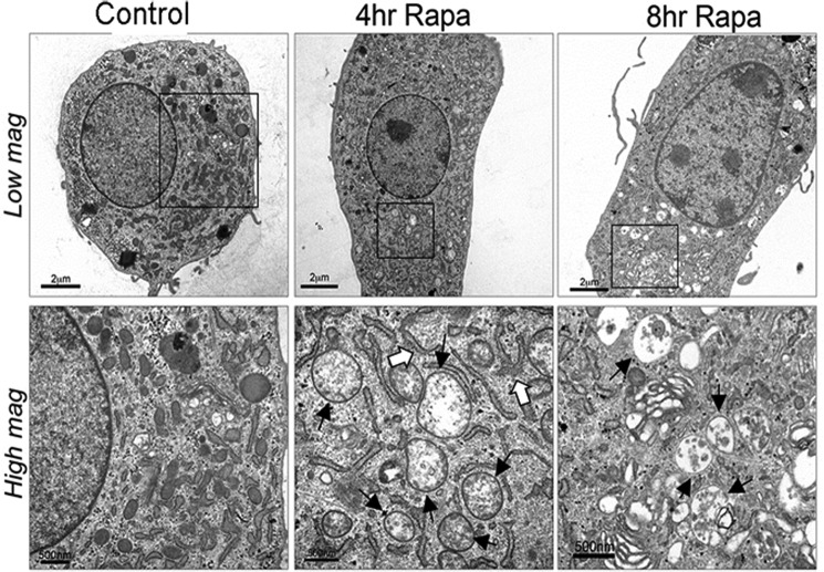
FIGURE 4.
Rapamcyin induces morphologic changes of increased autophagy by electron microscopy (EM). Porcine chondrocytes were incubated with or without rapamycin for 4–8 h. After fixation with glutaraldehyde, they were examined with TEM. In these representative images, typical autophagosomes (solid arrows) can be identified by the presence of double membranes and cytoplasmic components such as ribosomes and mitochondria and are visible at 4 and 8 h after rapamycin treatment. Pre-autophagosomal structures (open arrows), which are identified by their typical crescent shape, are present 4 h after rapamycin treatment.

