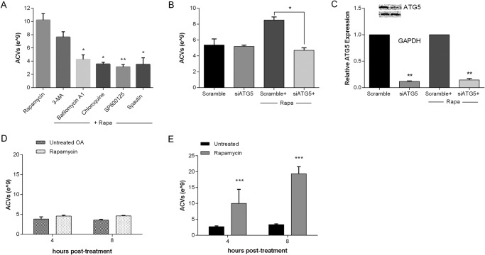
FIGURE 5.
Autophagy inhibition suppresses rapamycin-induced ACV production. A, porcine chondrocytes were incubated with no additives or autophagy inhibitors for 2 h, and then with rapamycin for an additional 2 h. ACVs were enumerated in conditioned medium. Values represent means ± standard deviations. All inhibitors tested except 3-MA significantly suppressed rapamycin-induced ACV numbers (n = 3, *, p < 0.05; **, p < 0.01). B, porcine chondrocytes were transfected with siATG5 or a scrambled control for 24 h. Medium was removed, and chondrocytes were exposed to no additives or to rapamycin for 8 h. ACVs in the conditioned medium were enumerated. Values represent means ± S.D. Cell treated with siATG5 produced less ACVs in the presence of rapamycin than those treated with scrambled control (n = 3, **, p < 0.01). C, RNA was isolated from chondrocytes treated with siATG5 or scrambled control. mRNA for ATG5 was quantified by real-time PCR. Values represent means ± S.D. siATG5 significantly reduced mRNA for ATG5 (n = 3, **, p < 0.01) and protein (inset). D, OA chondrocytes and E, normal human chondrocytes were treated with 50 μm rapamycin for 4–8 h. ACVs were enumerated. Values represent means ± S.D. Rapamycin did not increase ACV number from OA chondrocytes (n = 4, p > 0.05), but significantly increased ACV number in normal human chondrocytes (n = 3, ***, p < 0.001).

