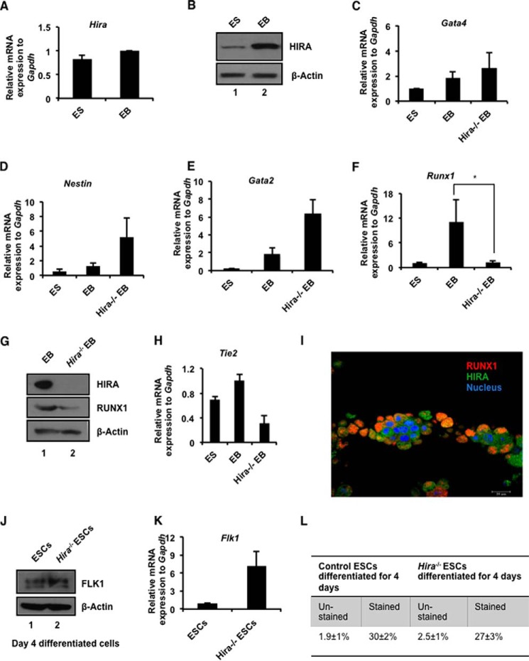
FIGURE 1.
HIRA influence RUNX1 in development. A, murine ES cells were differentiated into EBs in LIF free EB-specific medium and was subjected to RNA and protein isolation. Quantitative RT-PCR analysis of Hira in the ES-EB system (means ± S.E. for three independent experiments). B, the same sets of cells used in A were analyzed for the protein expression of HIRA by Western blot. C–F, control and Hira−/− ESCs were differentiated to EB formation. Quantitative RT-PCR analyses for different germ layer markers in ES/EB system (means ± S.E. for three independent experiments). F, the plot shows (p < 0.05) significant loss in expression of Runx1 in Hira−/−EB. G, Western blots for the same set of samples analyzed in C–F. H, quantitative RT-PCR analysis of Tie2 in ES-EB system (means ± S.E. for three independent experiments). I, mouse yolk sac was isolated at E9.5 and immunostained for RUNX1 and HIRA expression. Magnification was 63× under oil immersion. J, control and Hira−/− ESCs were differentiated toward mesodermal lineage by mesoderm inducer growth factors for the generation of HE. Cells at day 4 were analyzed for the expression of FLK1 at protein level. K, same set of cells used in J was analyzed for the expression of Flk1 at the mRNA level (means ± S.E. for three or more independent experiments). L, cells analyzed in J and K were sorted for FLK1 by FACS. The figure represents the fraction of FLK1+ control and Hira−/− cells. Averages of three independent experiments have been presented.

