FIGURE 6.
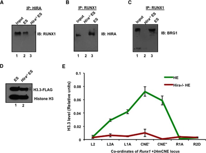
HIRA-mediated incorporation of histone H3.3 variant within Runx1 intronic enhancer drives hemogenic to hematopoietic transition. A and B, co-immunoprecipitation of HIRA and RUNX1 in control and Hira−/− ESCs. Shown are results of immunoprecipitation (IP) with HIRA and immunoblot (IB) with RUNX1 and vice versa, wherein input is 14%. C, co-immunoprecipitation of RUNX1 and BRG1 in control and Hira−/− ESCs. Shown are results of immunoprecipitation with RUNX1 and immunoblot with BRG1, wherein input is 14%. D, H3.3-Flag-tagged plasmid was transfected in ESCs by Lipofectamine and selected with G418 for 72 h. Western blot for FLAG-H3.3 in control and Hira−/− ESCs. E, HE cells were generated from transfected control and Hira−/− ESCs, cross-linked with 1% formaldehyde, and subjected to ChIP. A 3.6-kb locus flanking +24mCNE (conserved noncoding elements) region was analyzed to determine the extent of H3.3 incorporation. The plot shows the reduced or loss in incorporation of H3.3 in Hira−/− HE cells within +24mouse conserved noncoding elements (CNE) implicated in hemogenic to hematopoietic transition. PI is preimmune rabbit serum, used as negative control. Statistical analyses were performed using Student's t test function. *, p < 0.05; **, p < 0.01; ***, p < 0.001.

