FIGURE 3.
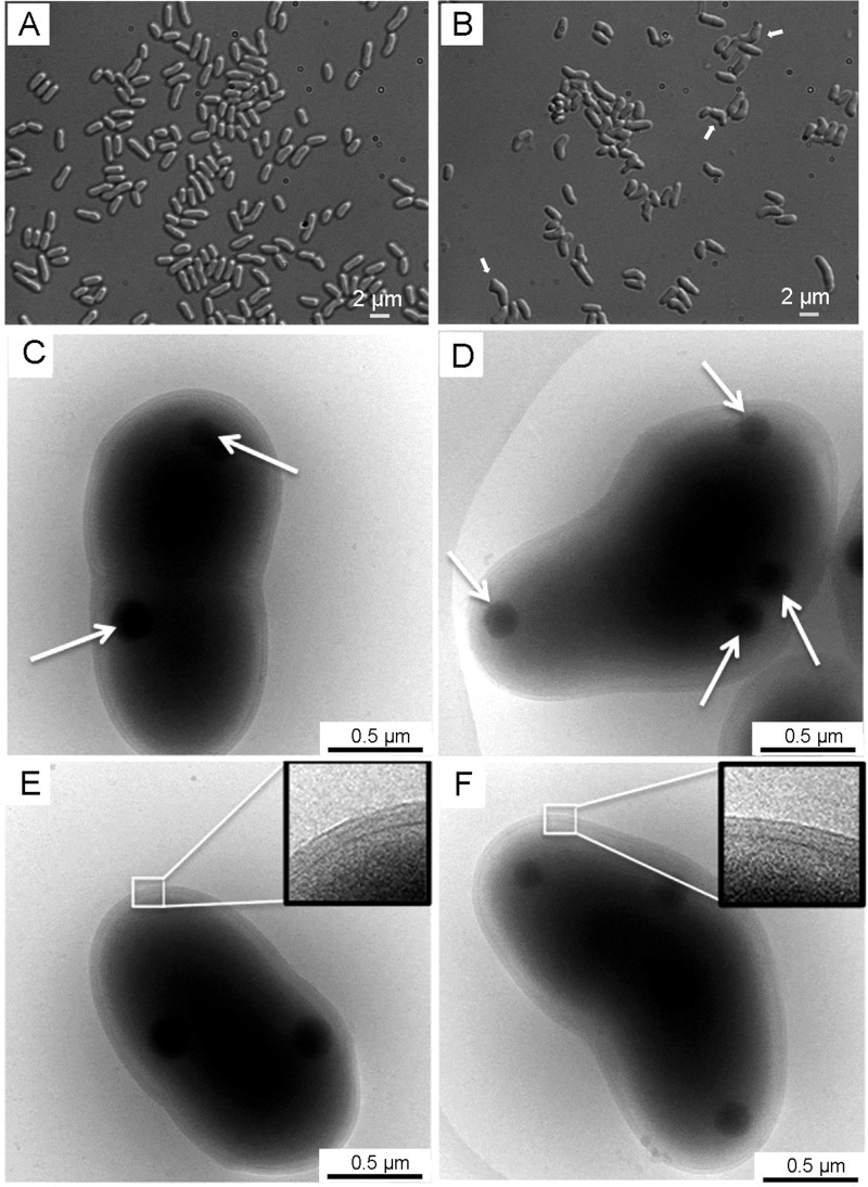
Morphologies of C. glutamicum strains. A and B, optical micrographs of exponentially growing C. glutamicum ATCC 13032 (A) and ΔltsA mutant (B) cells. Arrows indicate examples of cells exhibiting abnormal shapes. C–F, electron micrographs of frozen hydrated C. glutamicum ATCC 13032 cells (C and E) and of ΔltsA mutant cells exhibiting an irregular shape (D and F). The inset in E and F is an enlargement of the cell envelope. Arrows in C and D show the electron-dense granules.

