FIGURE 3.
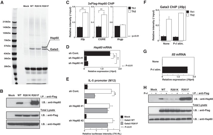
Hsp60 represses the transactivation of the Il5 promoter by methylated Gata3. A, total extracts from FLAG-Gata3 (WT or mutants)-expressing D10G4.1 cells were subjected to two-step affinity purification using a FLAG mAb and a Gata3 mAb, followed by SDS-PAGE and silver staining. The specific polypeptides were identified by mass spectrometry. Mock, mock-transfected. B, the FLAG-Gata3 (WT or mutants)-expressing D10G4.1 cells were subjected to an I.P. assay using a FLAG mAb, followed by immunoblotting (I.B.) with an Hsp60 Ab (upper). The total lysates were also subjected to I.B. in parallel (lower). C, naive CD4 T cells were stimulated under Th1 or Th2 conditions for 2 days and then infected with a retroviral vector carrying 3×FLAG-Hsp60 cDNA. Four days later, the binding of Hsp60 to the Il5 and Ifng promoters and conserved Gata3 response element (CGRE) region was determined by a ChIP assay using an anti-FLAG mAb. **, p < 0.01 by Student's t test. D, M12 cells were infected with a lentivirus encoding control (sh Cont.) or shHsp60 bicistronically with a puromycin resistance gene. The mRNA expression levels of Hsp60 were determined by RT-qPCR. **, p < 0.01 by Student's t test. E, reporter assays with the Il5 promoter were performed using Hsp60 knockdown M12 cells as in Fig. 2A. **, p < 0.01 by Student's t test. F, developing Th1 and Th2 cells were stimulated with or without phorbol 12-myristate 13-acetate (10 ng/ml) and ionomycin (500 nm) (P+I stim.) for 24 h. Then, the binding of Gata3 to the Il5 promoter was determined by a ChIP assay. G, the expression levels of Il5 mRNA were determined by RT-qPCR using a portion of the same cultured cells used in panel F. H, the FLAG-Gata3-expressing D10G4.1 cells were stimulated with or without phorbol 12-myristate 13-acetate (50 ng/ml) and ionomycin (500 nm) for 24 h. The amount of endogenous Hsp60 associated with the FLAG-tagged Gata3 was assessed by I.P., followed by I.B. (upper panel). The total lysates were also subjected to I.B. in parallel (lower panel).

