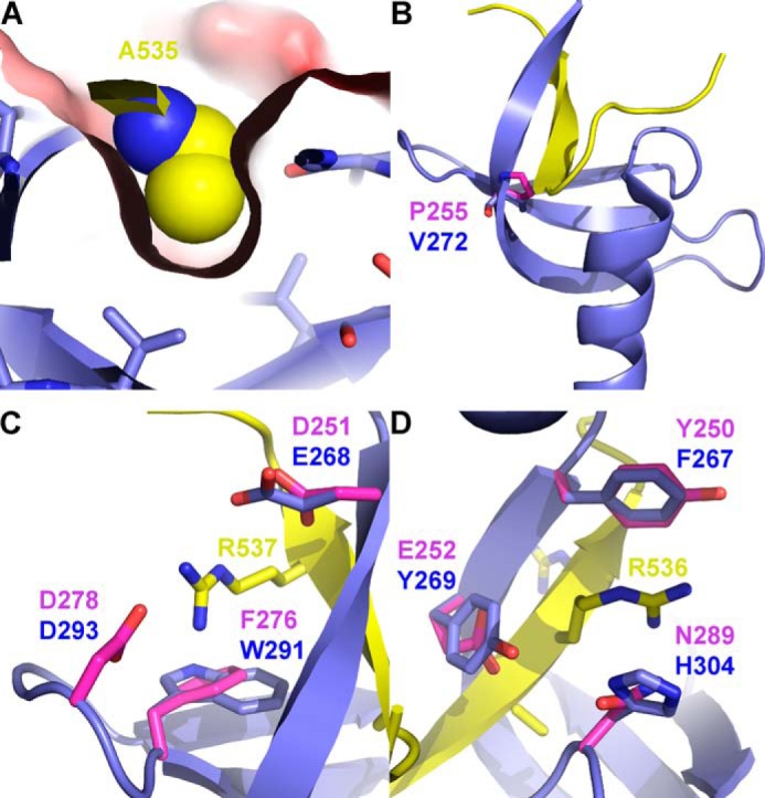
FIGURE 4.

Binding of Arabidopsis cpSRP54 by cpSRP43-CD2. A, the contact between Ala-535 of At-cpSRP54 (yellow) and At-cpSRP43-CD2 (blue) as observed in the complex structure (Protein Data Bank code 3UI2 (16)). Ala-535 is shown as space-filling spheres, whereas CD2 is shown as a stick model with the solvent-accessible surface colored by surface potential. B, the CD2 from At-cpSRP43 (blue) with the bound tail region of At-cpSRP54 (yellow) in a schematic representation. Val-272 of At-cpSRP43 is shown as sticks. In addition, Pro-255 from a homology model of Cr-cpSRP43 is shown as pink sticks. C and D, the twinned aromatic cages of cpSRP43. Superposition of the Arabidopsis complex structure of At-cpSRP43-ΔCD3 (blue) and the At-cpSRP54 tail region (yellow) (Protein Data Bank code 3UI2 (16)) with the homology model of Cr-cpSRP43 (pink) is shown. Residues forming the aromatic cages are shown as sticks and labeled accordingly (C, cage 1; D, cage 2). The two arginine residues (Arg-536 and Arg-537) from the At-cpSRP54 ARR motif are also shown as sticks.
