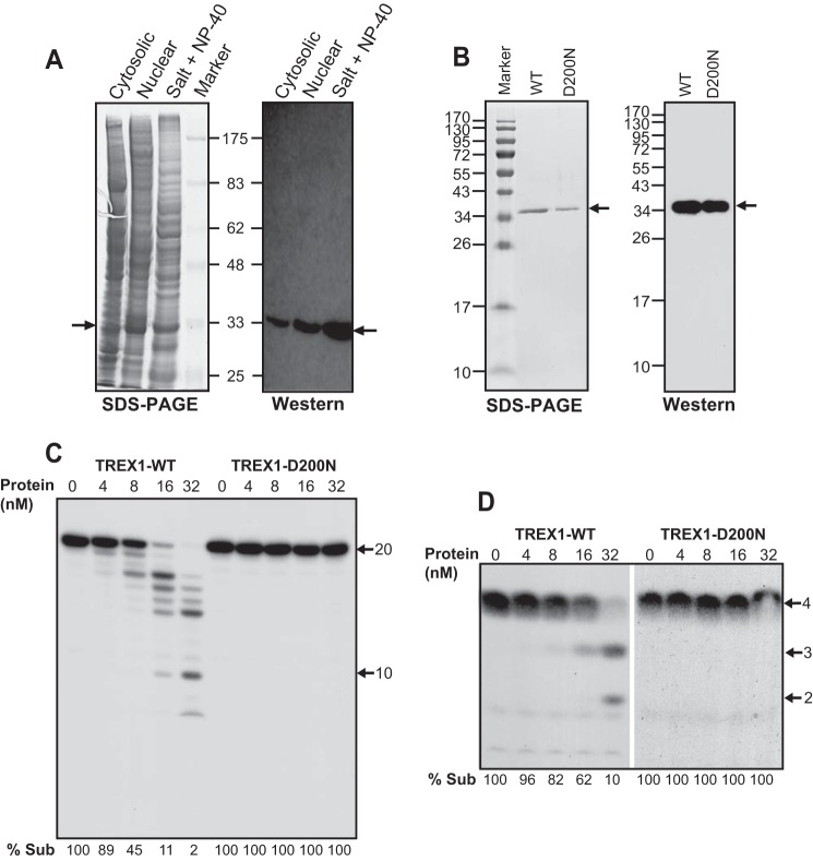
FIGURE 1.
Purified human TREX1 displays exoribonuclease activity on ssRNA. A, SDS-PAGE and Western blots of fractionated TREX1-containing extracts (40 μg each lane). Salt + Nonidet P-40 indicates the TREX1 protein samples after osmotic and detergent treatments. Arrows point to TREX1. Protein markers in kilodaltons are indicated. B, SDS-PAGE and Western analysis of the purified WT and D200N TREX1 proteins. The PAGE gel was stained with Coomassie Brilliant Blue R-250. Arrows point to the purified TREX1. Protein markers in kilodaltons are indicated. C, titration of TREX1-WT and TREX1-D200N proteins on a 20-nucleotide ssRNA substrate (2 nm) labeled at its 5′ end by [γ-32P]ATP using T4 polynucleotide kinase. Reaction time is 10 min. Arrows point to positions of the substrate and a 10-nt standard. % Sub indicates the percentage of the remaining substrate. D, activity of TREX1-WT and D200N on a 4-nucleotide poly(A) RNA substrate (2 nm) labeled at its 5′ end. Reaction time is 10 min. Arrows point to position of the 4-nt substrate and positions of the 2- and 3-nt standards.

